Objective
Electromyography refers to the procedure of extracting electrical signals from the human nervous system that flow through the various parts of the body and are intended to carry the information from the brain to the site of action. The objective of the work is to design a robust setup for efficient and accurate extraction of surface electromyographic signals that can be used to drive wearable devices as per human intent.
Research Aspects
- Data logging was done over Serial communication with a baud-rate of 57600 to collect over 2 million samples over 30 seconds of logging
- Post processing techniques included Principle Component Analysis (PCA), data re-filtering and data gap filling
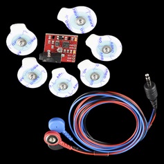
- Hardware signal were processed using high gain filter and noise attenuation circuits - Advancer Technologies Muscle Sensor V3
- GUI was capable of recording and analyzing 4 channels of electrodes simultaneously
- The software could provide real-time data across the globe
- GUI was developed over an open source platform - Processing
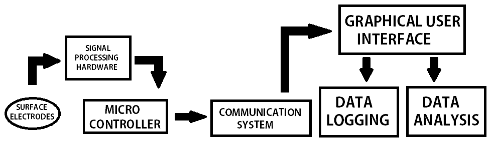
Flow Chart
Methodology
EMG signal extraction was performed using a dedicated electronic hardware. The hardware was supposed to extract the signals from the human skin using Ag-Cl surface electrodes. These signals are processed for amplification, noise reduction and filtering in order to be useful enough for study.
These signals were sent to an indigenously developed software for signal interpretation, data logging and analysis in real time. The pre-processing was data extraction involved skin preparation and electrode placement determination. The former was done by using Nuprep abrasive gel to get rid of the dead skin cells at the electrode site. To stregthen weak signals, Ten20 conductive paste was used.
Determing the correct muscles for electrode site was challenging as it is done solely by feel. Two electrodes are placed 0.5-1 inch apart at the terminals of the concerned muscle while the third electrode was placed at a neutral location. The resultant signal was the sum of two absolute differences between the MUAPs measured between the individual terminal electrodes with respect to the neutral electrode.
Results
- Efficiently extracted and recorded signals for more than 15 patients suffering from osteo-arthritis and analysed the results.
- Granted patent for the innovative design of cheap signal extraction setup
Gallery
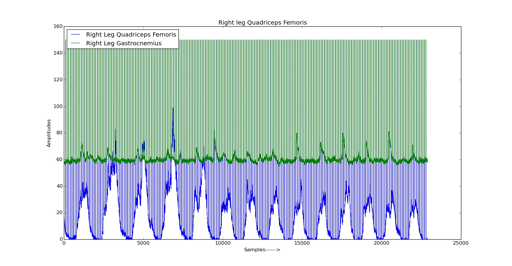
Raw Signals For Right Thigh Muscles
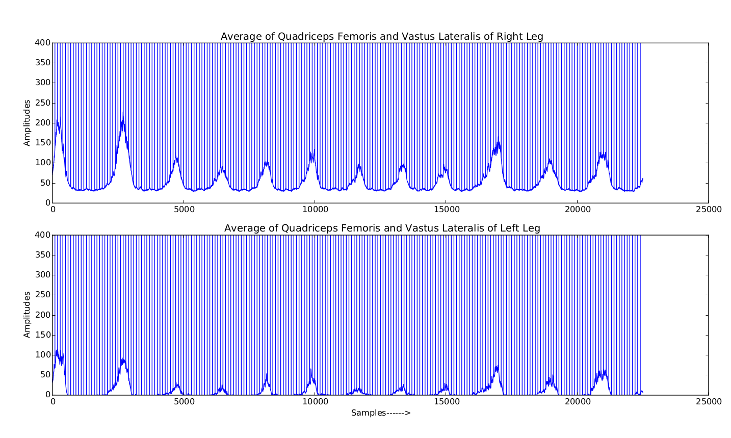
Average activation value of muscle activation suggesting patterns during flexion and extension of the knee

UI for the Data Logger